Optical spine and posture analysis with uEye industrial cameras
Back pain is one of the main causes of sick leave in Germany – often caused by lack of exercise, one-sided stress at work and being overweight. This is accompanied by strained muscles and ligaments and even wear and tear on the spine and intervertebral discs. Causes can also be osteoporosis, chronic inflammatory diseases or spinal deformities such as scoliosis. Innovative systems for 4D measurement of body statics can be used for precise diagnosis. DIERS International GmbH (Germany) enables fast and high-resolution optical measurement of the human back, spine and pelvis with DIERS formetric 4D – contactless and radiation-free. A dynamic version and additional modules for determining the mobility of the cervical spine or gait analysis as well as leg axis measurement make the system a compact movement analysis laboratory. Up to 5 camera units are integrated, equipped with uEye industrial cameras from IDS Imaging Development Systems GmbH.
Application
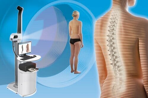
The DIERS formetric product family provides answers to a wide range of clinical questions concerning the objective and quantitative analysis of body statics and posture. Deformities such as scoliosis and all forms of spinal malformations can be visualised, and a wide variety of progress checks can be carried out. In this way, users such as orthopaedic surgeons can detect an imbalance in the muscular system or existing postural defects. The measurements are carried out statically, i.e. the patient stands in front of the system.
Back scan with the DIERS formetric 4D
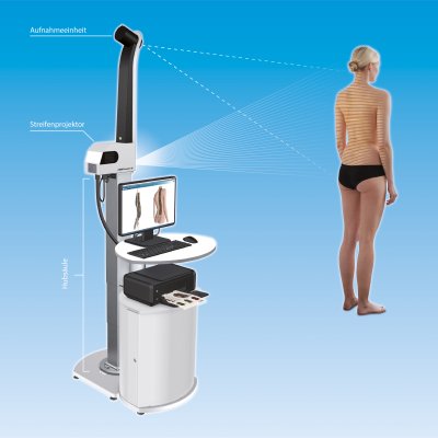
DIERS formetric surveying technology is physically based on the principle of triangulation. A light projector casts a grid of lines onto the patient’s back, which is recorded by a camera unit. Computer software analyses the line curvatures and generates a three-dimensional image of the surface using the method of photogrammetry (image measurement), similar to a virtual plaster cast. The examination is quick and absolutely radiation-free and contactless for the patient. This and the small space requirement of only 1.5 x 3 metres ensure high economic efficiency.
Compared to other systems, DIERS formetric provides not only a surface model of the back but also a 3D model of the spine without the use of reflective markers. It reconstructs the spatial course of the spine and the position of the pelvis independently. The 4D technology (3D + temporal component) additionally enables a very high accuracy and significantly increases the clinical informative value compared to 3D methods. By averaging a series of measurements, the influence of small body movements during the examination on the result can be minimised. In addition, measurement sequences are possible to carry out posture analyses and functional studies.
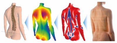
Spine surface, height map, spine curvature and strip projection (f.l.t.r.)
Additional components for a holistic movement analysis
In order to be able to carry out examinations in motion, the company also has a dynamic version of the DIERS formetric system in its range, DIERS 4D motion®. The patient runs on a treadmill. Using the latest projection and camera technology (60 images per second) and specially developed software, the complex interaction of the spine and pelvis can be measured during walking and displayed in moving images. This enables an analysis of the vertebral body rotation as well as the creation of movement models of the spine. Corrections such as spinal straightening or leg length compensation, e.g. through the use of shoe inserts, can be simulated.
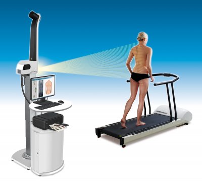
Dynamic spine measurement
Additional modules increase the range of applications up to a compact motion analysis laboratory. With the optional camera modules DIERS leg axis posterior and lateral for video-based analysis of the leg axis geometry, for example, asymmetries in stance as well as in the movement sequence can be determined and documented. The system software detects reflective markers applied to the patient and calculates movement patterns (dynamic) and angles (static and dynamic). In the case of foot and posture corrections, the effects on the leg axes can thus be directly displayed and checked.
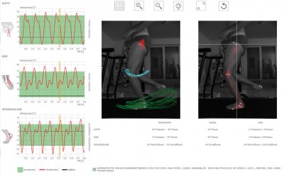
Video gait analysis and leg axis measurement
With the additional module “Cervical Spine”, the mobility of the cervical spine can also be recorded three-dimensionally. The measurement data and asymmetries are displayed graphically and can be analysed. The measurement process takes place using a special head hood. The DIERS formetric 4D system requires an additional camera system for the Cervical Spine Module.
As a high-end solution for holistic movement analysis at a running speed of up to 30 km/h, the DIERS 4D high performance Lab is also available to the user. The core technology here is also the DIERS formetric 4D back and spine measuring device, combined with 4 fast, powerful IDS cameras for video gait analysis of the legs from all sides and a treadmill with integrated foot pressure measuring plate. All devices are combined for simultaneous measurements of the whole body.
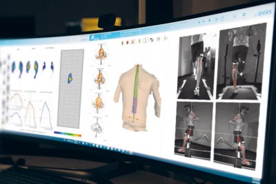
Holistic movement analysis at a running speed of up to 30 km/h
With the integration of these and other measurement systems, it is possible for the first time to view the complex biomechanical interaction in the human musculoskeletal system holistically. The spectrum of use ranges from medical diagnosis to training therapy and sports science. It enables the detection of scoliosis, pelvic obliquity and rotation, progress controls in osteoporosis and arthrosis, as well as neurological examinations (e.g. Romberg test), posture tests (Matthiass test, Flamingo test) and much more. Up to 5 uEye CP industrial cameras from IDS are used per unit. “We use the UI-3040CP as well as the UI-3240CP in our units. The USB 3 interface and the speeds achieved by both models convinced us,” explains Christian Diers, Managing Director/CEO of DIERS International GmbH.
Up to 5 uEye cameras per unit in use
The uEye CP stands for “Compact Power” and is suitable for all kinds of industrial applications thanks to its high level of functionality with comprehensive pixel pre-processing and its small size of only 29 x 29 x 29 mm. The internal 120 MB image memory for buffering image sequences also qualifies it for multi-camera systems. The USB 3.0 cameras enable a high data rate of 420 MByte/s, low CPU load and easy integration. Users can choose from a large number of modern CMOS sensors from manufacturers such as Sony, CMOSIS, e2v and ON Semiconductor with a wide range of resolutions.
The model UI-3040CP-M-GL Rev. 2 enables excellent image quality even in low light or when shooting fast-moving subjects. The integrated IMX273 global shutter CMOS sensor from Sony’s Pregius range scores particularly well for its image quality, high sensitivity and wide dynamic range. With a resolution of 1.57 MPixel (1448 x 1086 px) at 3.45 µm pixels, it achieves impressive 243 fps. This makes the camera ideal for classic industrial applications such as surface inspection, but also for detailed image evaluation in medical technology.
The UI-3240CP-M-GL Rev.2 is a particularly powerful industrial camera with the e2v 1.3 megapixel CMOS sensor. This sensor is one of the most sensitive sensors in the IDS portfolio. Besides its outstanding light sensitivity, the camera is distinguished by a number of additional functions: As an example, the sensor offers two global and rolling shutter variants that can be switched during operation, thus providing maximum flexibility for changing requirements and environmental conditions.
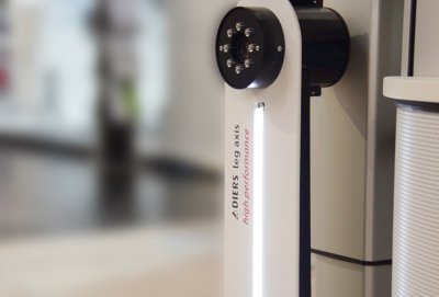
Camera module DIERS leg axis posterior high performance
Both models allow for easy integration into the customer application, as Christian Diers confirms: “We use the IDS SDK to integrate the cameras into our software. The added value for us lies in the simple verification of the correct functioning and integration of the cameras into our system. Environmental settings, such as brightness or contrast, are controlled by the IDS software package.”
Outlook
The medical technology market is highly regulated, so that changes and new developments of products must be well considered through elaborate certification and approval procedures. At the same time, price pressure is increasing. Nevertheless, DIERS is currently working on various new developments – from insole production to research projects in the rehabilitation and care sector. In addition to the use of classic 2D industrial cameras, 3D models are also conceivable, which inherently introduce the spatial component.
Medical technology is an exciting environment for research and development, as it often creates new hope and perspectives for patients with new products. Image processing has a wide range of applications in this field and can hold the potential for completely new analysis and diagnostic procedures – absolutely contactless, radiation-free and thus gentle and painless for those being treated. Like here at DIERS international: Vertebrae by vertebrae, picture by picture to the application with perspective.
Client
DIERS International GmbH is an innovative, family-run company that has been successfully developing, manufacturing and distributing biomechanical measuring systems since 1996. The aim here is to meet the increasing clinical requirements of interdisciplinary medicine. The company thus offers a wide range of systems for measuring the human musculoskeletal system.
www.diers.com

